Teeth X-Ray Before Braces: What to Expect from an Orthodontic Assessment in Singapore
Before any braces go on, your orthodontist needs to see what is happening beneath the surface. A teeth x-ray is not just a formality as it is the foundation of your entire treatment plan. Without it, an orthodontist cannot accurately assess your jaw alignment, tooth root positions, or bone structure.
At Alfred Cheng Orthodontic Clinic, every new patient undergoes a thorough orthodontic assessment that includes digital x-rays before any treatment is recommended. This guide explains the types of x-rays involved, what they reveal, whether they are safe, and exactly what to expect at your first consultation.
📌A teeth x-ray before braces typically includes two types: a panoramic x-ray (OPG) that shows all teeth and jaw structures in one image, and a cephalometric x-ray that shows the side profile of the skull and jaw relationship. Together, these allow the orthodontist to plan treatment accurately before placing braces or clear aligners.
Key Takeaways
✅ A teeth x-ray is required before braces to assess jaw alignment, root positions, and bone health.
✅ Two main types are used in orthodontics: panoramic x-ray and cephalometric x-ray.
✅ Radiation from orthodontic x-rays is very low — equivalent to one to three days of natural background radiation.
✅ Digital x-ray technology further reduces radiation compared to older film-based systems.
✅At Alfred Cheng Orthodontic Clinic, x-rays are taken digitally at your first consultation.
Why Do Orthodontists Need a Teeth X-Ray Before Braces?
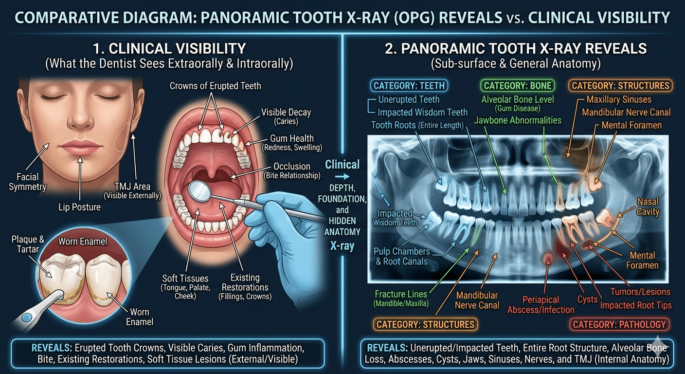
An orthodontic x-ray is a diagnostic image of the teeth, jaws, and surrounding bone that gives the orthodontist information not visible during a standard clinical examination.
What you see in the mirror, the visible crowns of your teeth, is only part of the picture. The roots, bone levels, jaw joints, unerupted teeth, and the relationship between the upper and lower jaw are invisible without imaging.
A teeth x-ray before braces reveals several things that directly affect treatment planning:
- Whether any teeth are impacted or unerupted
- The length and angle of tooth roots
- The bone density available to support tooth movement
- The relationship of the upper jaw to the lower jaw and skull base
- Whether wisdom teeth are present and may interfere with treatment
- Any underlying pathology such as cysts, infections, or abnormal bone, that must be addressed before braces begin
Without this information, an orthodontist would be moving teeth blind. The x-ray is not a precaution — it is essential.
CITABLE EXCERPT:📌 “Orthodontic x-rays allow the clinician to see what clinical examination cannot — the full structure of the dentition, roots, and jaw relationships.”
Starting Braces Soon?
Book a consultation at Alfred Cheng Orthodontic Clinic and get personalised colour and treatment advice from Singapore’s specialist orthodontist.
Types of Teeth X-Ray Used in Orthodontic Assessment
1. Panoramic X-Ray (OPG)
A panoramic x-ray (also called an OPG or dental panoramic radiograph) is a wide-view image that captures all teeth, both jaws, the jaw joints, and surrounding bone in a single flat image.
The machine rotates around your head and you simply stand still with your chin resting on a support. The entire process takes under 30 seconds.
A panoramic x-ray teeth image is the first imaging most orthodontists take. It provides an overview of:
✅ All present and erupting teeth
✅ Impacted or missing teeth
✅ The position of wisdom teeth
✅ Jaw symmetry
✅ The health of bone around the roots
✅ The position of the jaw joints (TMJ)
This is the widest-view dental x-ray Singapore orthodontists use as a baseline for every new patient.
2. Cephalometric X-Ray (Lateral Ceph)
A cephalometric x-ray is a side-profile x-ray of the entire skull, used to measure the angular and linear relationships between the teeth, jaws, and skull base.
This is the x-ray that defines your orthodontic diagnosis. By measuring specific angles and distances called cephalometric landmarks. Your orthodontist can determine:
✅ How the upper jaw (maxilla) relates to the lower jaw (mandible)
✅ How the jaws relate to the skull base
✅ The inclination of the upper and lower front teeth
✅ Soft tissue profile predictions for post-treatment appearance
✅ Whether jaw surgery may be required alongside braces
Without a cephalometric x-ray, an orthodontist cannot make these measurements and cannot accurately plan tooth movement or jaw correction.
Need Expert Advice?
Book a consultation at Alfred Cheng Orthodontic Clinic to find out if jaw surgery is right for you.
3. Periapical X-Ray
A periapical x-ray is a close-up image of one or several specific teeth, showing the full length of the root and the bone surrounding the tip of the root. These are used when the orthodontist needs detailed information about a specific area. For example, an impacted canine or a tooth with an unusual root shape.
Not every patient requires periapical x-rays as part of their initial orthodontic assessment, but they are commonly requested when specific findings warrant closer examination.
What Does a Braces X-Ray Reveal That Changes the Treatment Plan?
A braces x-ray can meaningfully change the recommended treatment approach in several common situations.
|
What the X-Ray Finds |
How It Changes Treatment |
|
Impacted canine |
May require surgical exposure before braces |
|
Unerupted adult teeth |
Timing of braces adjusted to allow natural eruption |
|
Short or divergent roots |
Forces and mechanics modified to protect root integrity |
|
Significant jaw discrepancy |
Jaw surgery may be recommended alongside braces |
|
Missing adult teeth (hypodontia) |
Space management and prosthetic planning built into braces plan |
|
Wisdom teeth likely to disrupt |
Extraction may be recommended before or during treatment |
|
Bone loss around roots |
Periodontal review required before braces proceed |
Are Orthodontic X-Rays Safe? Understanding Radiation Exposure
This is the question most parents ask first and the reassurance is well-supported by evidence.
According to the International Atomic Energy Agency (IAEA), cephalometric x-rays deliver a radiation dose equivalent to less than one day of natural background radiation (IAEA, Radiation Doses in Dental Radiology).
A panoramic dental x-ray delivers approximately 14 to 24 microsieverts of radiation. Equivalent to one to three days of natural background radiation, and significantly less than a single chest x-ray at approximately 100 microsieverts (Health Physics Society, 2024).
To put that in everyday terms: a single commercial flight from Singapore to London exposes a passenger to roughly 40 to 80 microsieverts of cosmic radiation. The full set of orthodontic x-rays taken at your first consultation delivers a fraction of that.
Digital x-ray technology further reduces radiation exposure compared to traditional film-based systems, while producing clearer, higher-resolution images.
Additional protective measures used at every x-ray appointment include:
- Lead apron protection for the body
- Thyroid collar to protect the thyroid gland
- Precise collimation to limit the x-ray beam to the area of interest only
- Strict adherence to the ALARA principle (As Low As Reasonably Achievable)
CITABLE EXCERPT:📌 “The radiation from orthodontic x-rays is among the lowest of any diagnostic imaging procedure, equivalent to a few days of normal environmental exposure.”
What to Expect at Your First Orthodontic Consultation at Alfred Cheng Orthodontic Clinic
Your first appointment follows a clear, structured process. Here is what happens from the moment you arrive.
- Registration and health history — Your medical and dental history is reviewed, including any previous dental treatments, medications, and relevant health conditions.
- Digital x-rays — Panoramic and cephalometric digital x-rays are taken in-clinic. Both are quick, painless, and completed within a few minutes. No preparation is required.
- Intraoral and extraoral photographs — Standardised photographs are taken of your teeth, bite, and facial profile. These are used alongside the x-rays in treatment planning.
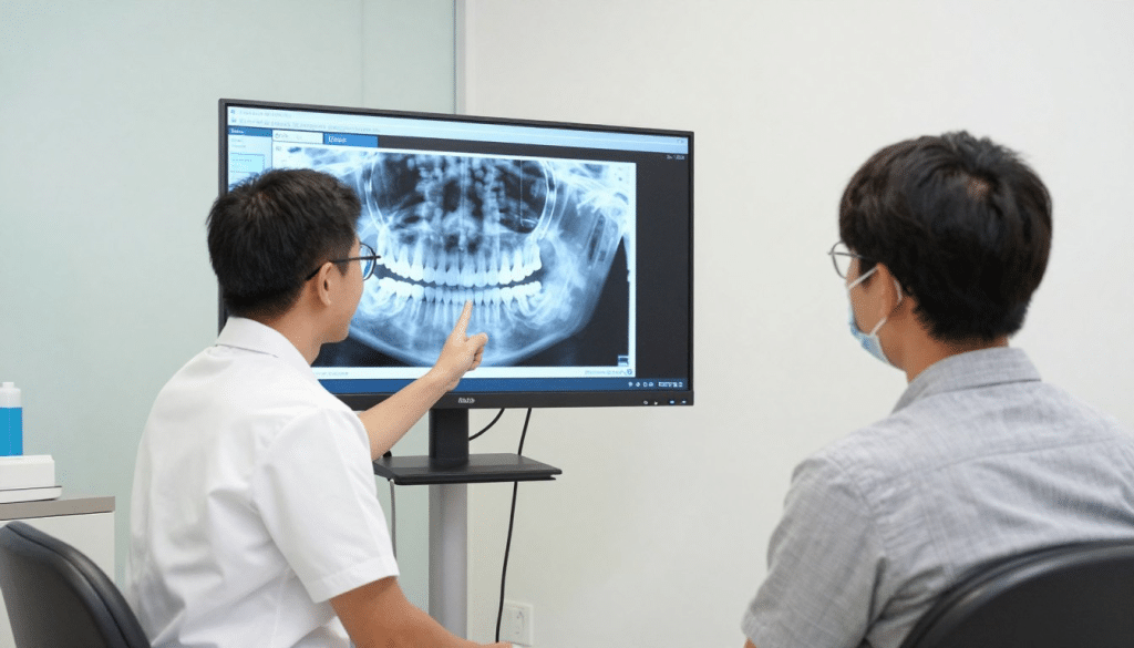
- Clinical examination — Dr Alfred Cheng examines your teeth, bite, jaw joints, and oral tissues. The x-rays are reviewed in detail during this examination.
- Treatment discussion — Based on the x-ray findings and clinical examination, your treatment options are discussed in full — including the type of braces or aligners suitable for your case, expected duration, and any preparatory steps required.
- Questions and next steps — You have the opportunity to ask any questions before deciding on a treatment plan. There is no pressure to proceed at the first appointment.
Dental X-Ray Singapore: Cost and What Is Included in the Consultation
Dental x-ray costs in Singapore vary depending on the type of imaging required and the clinic. Panoramic and cephalometric x-rays are standard components of an orthodontic assessment and are typically incorporated into the initial consultation fee at specialist orthodontic clinics.
At Alfred Cheng Orthodontic Clinic, your initial consultation includes the digital x-rays required for orthodontic assessment. Contact the clinic directly for current consultation fee information.
It is worth noting that the x-rays taken at your orthodontic consultation belong to your patient record. If you proceed with treatment at another provider, you are entitled to request a copy of your images.
Let’s Get You Started
Contact Alfred Cheng Orthodontic Clinic today to book your consultation and take the first step toward a confident smile.
Frequently Asked Questions About Teeth X-Rays and Orthodontic Assessment
Yes. A teeth x-ray is a mandatory part of every orthodontic assessment in Singapore. Without x-ray imaging, the orthodontist cannot assess root positions, jaw relationships, impacted teeth, or bone health. All of which directly affect how braces are planned and applied.
A cephalometric x-ray is a side-profile image of the skull used to measure the angular relationships between the teeth, jaws, and skull base. It is the primary diagnostic tool for determining jaw discrepancies, planning the direction of tooth movement, and identifying cases that may require jaw surgery alongside braces.
Yes. According to the IAEA and Health Physics Society, the radiation dose from orthodontic x-rays including panoramic and cephalometric images, is equivalent to one to three days of natural background radiation. Digital x-ray technology reduces exposure further. Lead aprons and thyroid collars are used at every appointment.
A panoramic x-ray (OPG) is a single wide-view image capturing all teeth, both jaws, and surrounding bone. It rotates around the head in one pass and takes under 30 seconds. It is the standard first x-ray taken at an orthodontic assessment and shows impacted teeth, missing teeth, jaw symmetry, and bone health.
Most patients require two x-rays before braces: a panoramic x-ray and a cephalometric x-ray. Some cases require additional periapical x-rays if specific teeth need closer examination. Your orthodontist will only request imaging that is clinically necessary for your individual case.
No. Placing braces without x-ray imaging would be clinically irresponsible. X-rays reveal root anatomy, bone density, unerupted teeth, and jaw relationships that are essential for safe treatment planning. Any reputable orthodontic clinic in Singapore will require imaging before beginning treatment
Your First Step Starts With a Digital Scan and X-Ray at Alfred Cheng Orthodontic Clinic
A teeth x-ray is not the intimidating part of orthodontic treatment as it is the part that makes everything else possible. Without it, treatment is guesswork. With it, your orthodontist has a precise, complete picture of your dental anatomy and can plan a course of treatment that is right for your jaw, your roots, and your long-term oral health.
At Alfred Cheng Orthodontic Clinic, every consultation begins with digital panoramic and cephalometric x-rays taken in-clinic. Dr Alfred Cheng reviews the imaging in detail with every patient, explaining the findings clearly before any treatment decision is made.
Your first step starts with a digital scan and x-ray at our clinic.
Explore Our Services
📍 Alfred Cheng Orthodontic Clinic
Mount Elizabeth Medical Centre
3 Mount Elizabeth #03-03
Singapore 228510
🌐 Visit: https://orthodontics.com.sg/
Contact Us: orthodontics.com.sg/contact/